Uncovering the Brain’s Flexible Mechanisms for Representing Diverse Numbers

– Providing evidence for the hierarchical representation of relative numerical magnitudes in the human brain –
March 25, 2025

National Institute of Information and Communications Technology

Abstract

The National Institute of Information and Communications Technology (NICT, President: TOKUDA Hideyuki, Ph.D.) has revealed, through fMRI-based brain activity analysis, that multiple regions in the human cerebral cortex flexibly represent numerical quantity. This finding comes from research by HAYASHI Masamichi (Researcher (Tenure-Track)) at Center for Information and Neural Networks (CiNet), part of NICT’s Advanced ICT Research Institute, in collaboration with the University of Tokyo’s graduate student KIDO Teruaki (NICT cooperative visiting researcher), and Prof. YOTSUMOTO Yuko.
Although certain brain areas are known to respond to numerical quantity, this study expands that understanding by showing that some regions respond to relative quantity (e.g., “extra-small,” “small,” “large,” and “extra-large”) rather than absolute quantity (i.e., specific quantity). Moreover, these context-dependent, relative representations become more pronounced along the pathway from the parietal to the frontal lobe.
These results highlight the flexible nature of numerical quantity processing in the brain, and they are expected to advance our understanding of how the brain handles other types of “magnitude” concepts, including time and size.
This work was published in the journal “Nature Communications” on January 6, 2025.

Background

It has long been known that certain brain regions contain neurons that respond selectively to specific numbers. However, it remained unclear whether they always react to the same numbers, such as 8 or 15, regardless of context (absolute representation), or if their response changes depending on the situation. If neurons strictly followed absolute quantity, an impractically large number of neurons would be required to process infinite numerical quantities, making it unclear how the human brain, with its limited neurons, efficiently processes such a wide range of numbers.

Achievements

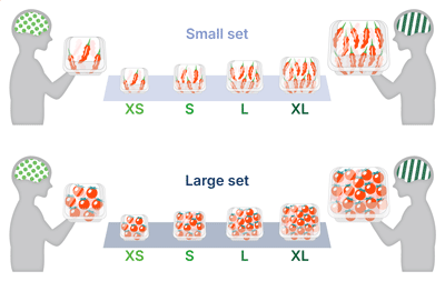
Figure 1 Relative representation of numerical quantity in the brain Although the actual number of items differs, the brain exhibits similar activity patterns when focusing on an extra-small (XS) quantity among packs with few items (small set) and when focusing on an XS quantity among packs with many items (large set). Similarly, regardless of the set, focusing on an extra-large (XL) quantity produces a comparable pattern of brain activity. [Click picture to enlarge]
In this study, we used functional magnetic resonance imaging (fMRI) to measure participants’ brain activity and analyzed it with multivariate pattern analysis. Over three days, participants viewed black-and-white dot patterns that presented numerical information in different ranges while their brain activity was recorded.
The results showed that despite variations in numbers, certain brain regions exhibited similar activity patterns (Figure 1). This occurred when the relative magnitude within the range was the same (e.g., XS in the large set and that in the small set). This suggests that neurons may adjust their responses to numbers based on context (i.e., the numerical range), enabling efficient encoding while conserving neural resources.
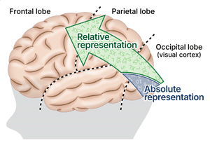
Furthermore, our study revealed a hierarchical structure in visual processing: lower sensory regions represented numbers in absolute terms, whereas higher-order cortices, from the parietal to the frontal lobe, gradually shifted toward relative numerical quantity representations. This shift highlights how the brain flexibly encodes numerical magnitude based on context (Figure 2).
Numerical information is embedded in various forms of media, and its accurate and effective communication plays a crucial role in determining the quality of communication. This study aims to enhance communication quality by uncovering the brain functions involved in processing numerical concepts.
Figure 2 Hierarchy of absolute and relative representations of numerical quantity
[Click picture to enlarge]

Future Prospects

While this study focused on numerical quantity, similar mechanisms may also underlie other quantitative concepts, such as size and time. Investigating whether the brain represents these concepts in a relative manner could provide deeper insights into how the brain perceives and interprets its environment as a whole.

Article information

Journal: Nature Communications
DOI: 10.1038/s41467-024-55599-8
Title: Hierarchical representations of relative numerical magnitudes in the human frontoparietal cortex
Authors: Teruaki Kido, Yuko Yotsumoto, Masamichi J. Hayashi

Research team

KIDO Teruaki
Graduate Student, Department of Life Sciences, Graduate School of Arts and Sciences, The University of Tokyo, Tokyo, Japan
Cooperative Visiting Researcher, Center for Information and Neural Networks (CiNet), Advanced ICT Research Institute, National Institute of Information and Communications Technology, Suita, Japan

YOTSUMOTO Yuko
Professor, Department of Life Sciences, Graduate School of Arts and Sciences, The University of Tokyo, Tokyo, Japan

HAYASHI Masamichi
Researcher (Tenure-Track), Center for Information and Neural Networks (CiNet), Advanced ICT Research Institute, National Institute of Information and Communications Technology, Suita, Japan
Guest Associate Professor, Graduate School of Frontier Biosciences, Osaka University, Suita, Japan
Appendix

Experimental Procedure

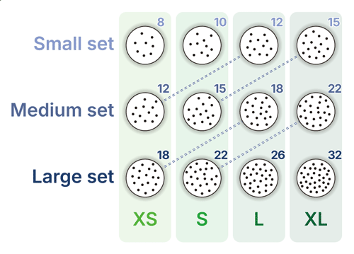
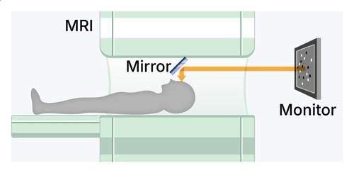
In this study, we measured brain activity using fMRI while participants viewed dot patterns and focused on the number of dots (Figure 3). The dot patterns were divided into three sets: 8, 10, 12, and 15 dots (small set), 12, 15, 18, and 22 dots (medium set), and 18, 22, 26, and 32 dots (large set). Although the absolute numbers varied, each set was categorized into four relative levels: “XS,” “S,” “L” and “XL” (Figure 4). 
To examine how the brain processes relative numerical magnitudes, each dot pattern set was presented on a different experimental day. The results showed that certain brain regions exhibited similar activity patterns for the same relative category within a set, regardless of the actual number of dots. In other words, specific brain areas consistently responded similarly to dot patterns categorized as “XS,” “S,” “L” and “XL” regardless of the absolute number of dots.
Figure 3 Experimental setup
Figure 4 Dot pattern sets

Glossary

Functional magnetic resonance imaging (fMRI) When neurons in the brain become active, they require more oxygen, leading to an increase in the proportion of oxygenated hemoglobin, which is a form of hemoglobin in red blood cells that is bound to oxygen. fMRI is a specialized imaging technique that utilizes MRI to detect this as a change of signal intensity known as the blood-oxygen-level-dependent (BOLD) signal. In fMRI, the brain is divided into millimeter-scale cubic units called voxels, and BOLD signals are measured for each voxel. This allows researchers to identify which areas of the brain are active. Back to contents

Multivariate pattern analysis (MVPA) Multivariate Pattern Analysis (MVPA) is a method to decode how the brain represents information by analyzing the pattern of BOLD signals across voxels. Back to contents

Technical Contact

HAYASHI Masamichi
Brain Networks and Communication Laboratory,
Center for Information and Neural Networks,
Advanced ICT Research Institute

Media Contact

Press Office
Public Relations Department